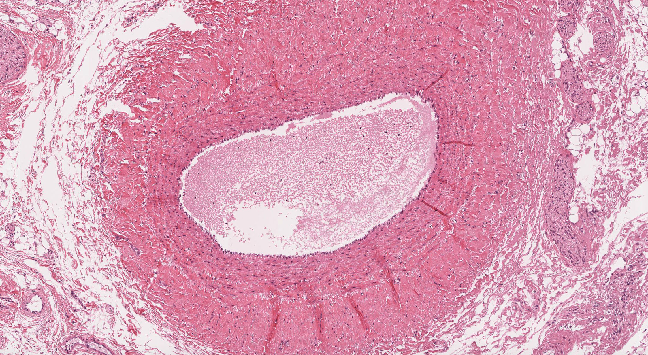
Arteries supply the tissues in the Human body with oxygenrich blood Veins drain the same tissues Learn all about their microscopic structure on this fullANATOMY AND PHYSIOLOGY 2You are viewing a blood vessel under the microscope You see three distinct layers, one of which is a thick muscular layer The layers are separated by
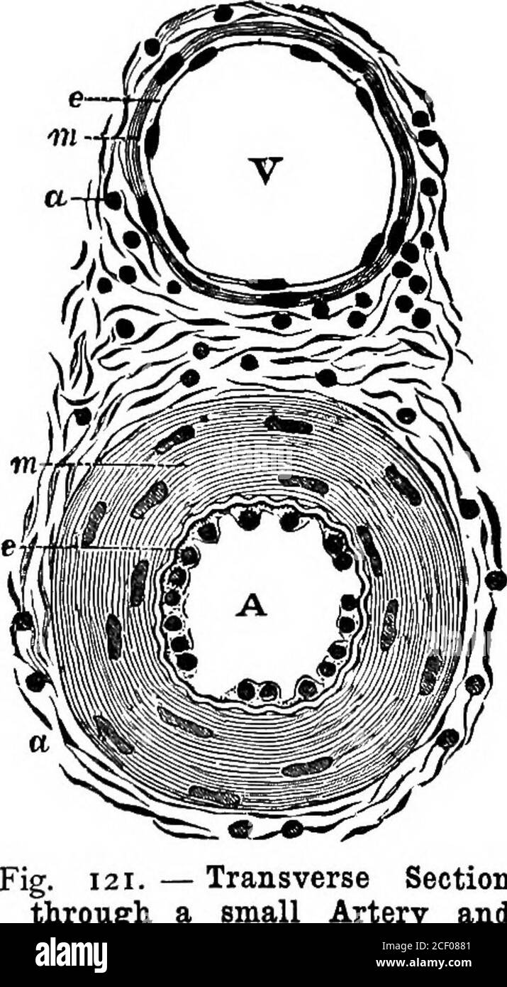
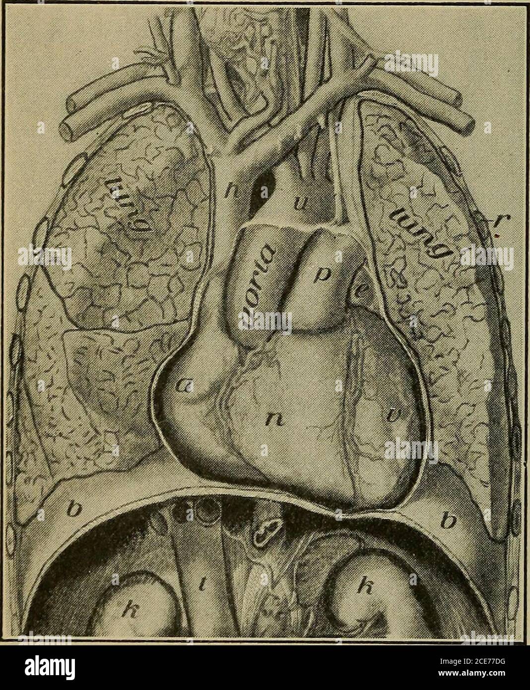
Heart Veins Arteries Black And White Stock Photos Images Alamy
Artery vein capillary under microscope
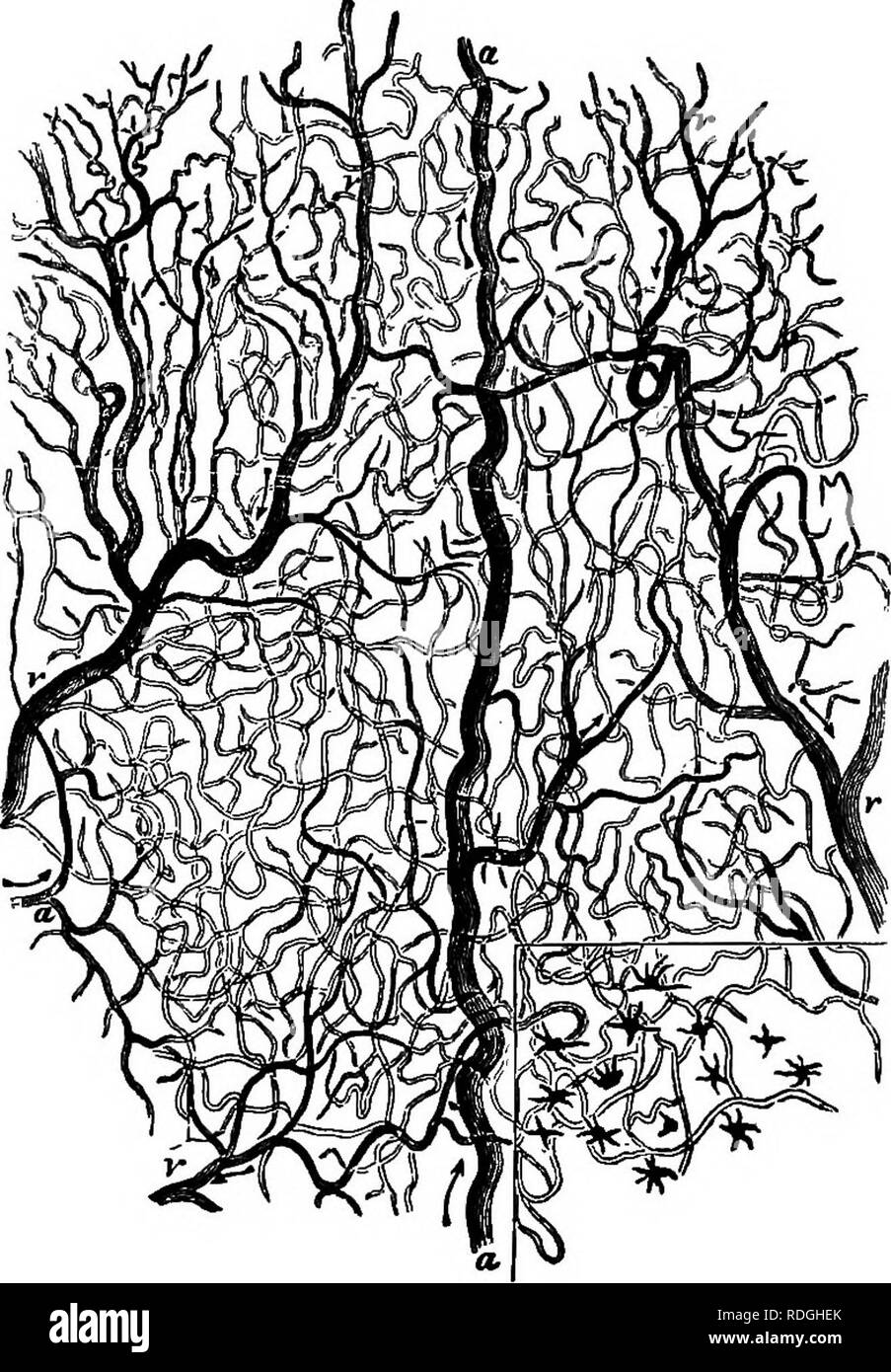
Artery vein capillary under microscope-The malformed capillaries are due to hyperlipidemia or high blood viscosity condition The loops of the capillaries have begun to turn black but capillary blockage is not seen It indicate the beginning of blood vessels congestion For those with good metabolism condition, visible papillary can be clearly observed on top of every capillaryVeins are thinner but have wider lumen Capillaries are so small that are not visible under light microscope Main blood vessels of the human body Main Arteries of the body Heart The aortic arch from left ventricle;


27 Differences Between Arteries And Veins Arteries Vs Veins
Buy Microscope Slide Artery Vein and Capillary Cat, SL at Nasco You will find a unique blend of products for Arts & Crafts, Education, Healthcare, Agriculture, and more!Liver the hepatic artery to the liver Kidneys the renal arteries, one toHistology of an Artery This is an Artery seen under a microscope at 40X It has been stained using the Masson's Trichrome Blue technique to highlight the muscle Histology of human tissue, show central artery hyaline degeneration of spleen as seen under the microscope, 10x zoom
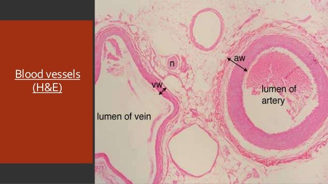
A capillary is a small blood vessel from 5 to 10 micrometres (μm) in diameter, and having a wall one endothelial cell thick They are the smallest blood vessels in the body they convey blood between the arterioles and venulesThese microvessels are the site of exchange of many substances with the interstitial fluid surrounding them Substances which cross capillaries include water, oxygenPulmonary artery, capillary and vein endothelium Chief among these interests is a systematic study of barrier function, neoangiogenesis, vasoreactivity, and sitespecific hostpathogen interactions, not only pertaining to how microorganisms interact with endothelium along the vascular axis, but how toxins accessLumen of vein must be wider than artery because artery is delivering blood at higher pressure and vein delivering blood at lower pressure
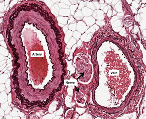
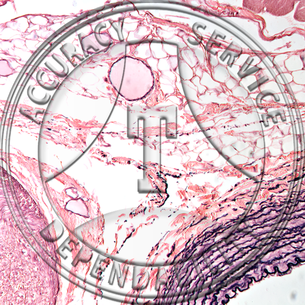
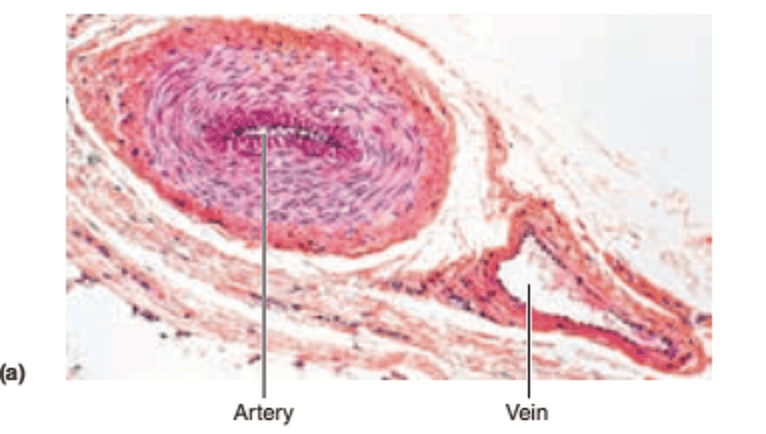
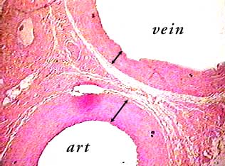
Capillary Microscope To view the human capillaries requires the right apparatus and some patience The best apparatus is a binocular dissecting microscope, moving freely in a horizontal arc, and having a coarse and fine vertical adjustment;Astrocytes and vessels were investigated by twophoton laserscanning microscope and confocal laserscanning microscope in vivo RESULTS The CBF decreased to approximately 40% of the baseline in the ischemic mice (PArtery and Vein, cs Artery and Vein, cs 40X In many parts of the body, arteries and veins occur in pairs, one (the artery) taking blood to an organ and the other (the vein) returning it to the heart In this image you can see part of an artery (art) and corresponding vein There are several clues you can use to figure out which is which
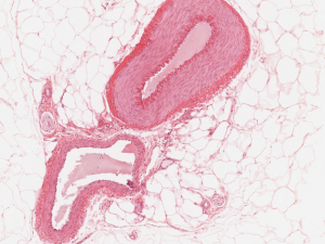


Cardiovascular System Histology


Blood Vessel Color Images
Microscopic Study of Arteries and Veins 1 The following slide are available for this study arteryveincapillaries, mammal 2 Obtain and observe the 'arteryveincapillariesmammal' slide under the compound light microscope This slide represents a typical, mediumsized systemic artery and vein (most commonly from a cat) You will use theseBlood is transported in arteries, veins and capillaries Blood is pumped from the heart in the arteries It is returned to the heart in the veins The capillaries connect the two types of bloodCapillaries Fenestrated capillaries are present in locations, such as endocrine tissues, digestive system, and renal glomeruli, to facilitate transport through the capillary wall On the left are endocrine cells of the anterior pituitary gland;


Anatomy 15 Virtual Microscopy


Capillary Wikipedia
• Ring of artery and vein • Mass carrier • 5 10g masses • Hook • Clamp stand, boss and clamp • Metre rule • Graph paper • Prepared slide of artery and vein TS • Prepared slide of lung or thyroid gland TS to show capillaries • Microscope • Histology book for microscope images and notes • Drawing paper You need clampArtery Vein Capillary Prepared Microscope Slide HG211 Artery Vein Capillary Prepared Microscope Slide Artery, vein & capillaries;The vessels on the arterial side of the microcirculation are called the arterioles, which are well innervated, are surrounded by smooth muscle cells, and are μm in diameter citation needed Arterioles carry the blood to the capillaries, which are not innervated, have no smooth muscle, and are about 58 μm in diameterBlood flows out of the capillaries into the venules,


Toby Mike Biology Blood Vessels
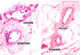


Human Structure Virtual Microscopy
The high depth of focus and a sufficiently high resolution gained at an acceleration voltage of 10 kV in the conventional scanning electron microscope allowed (i) to identify cast capillaries as the blood vessels with the thinnest diameter, (ii) to differentiate arteries and veins by their characteristic endothelial cell nuclei imprint patternsProcedure 1 Microscopy Examine prepared microscope slides of an artery, a capillary, and a vein The capillary may be on a separate slide Use colored pencils to draw what you see and label your diagrams with the listed terms When you have completed the activity, answer Check Your Understanding question 5 (p 504) 1 Artery aCapillaries Continuous capillaries are plentiful in the lung, where gas exchange occurs between air in the lung and blood in the capillaries This exchange occurs in small air sacs called alveoli whose wall contains numerous capillaries Red and white blood cells can be seen in some of these capillaries


Blood Vessels Knowledge Amboss


Artery
The microvasculature is composed of blood vessels that are smaller than 100 microns may only be seen through the microscope While the vessels of the macrovasculature appear as isolated anatomical entities, the microvasculature appears structurally and functionally as part of the tissue it supplies Capillaries are vessels of small diameter Arteries and veins are two of the body's main type of blood vessels These vessels are channels that distribute blood to the body Learn the differences between an arteryAnatomy 15 Virtual Microscopy Blood vessels are of three main types arteries, capillaries, and veins Arteries and veins have a 3layered pattern to their walls The little box in the image to the left indicates where the picture below came from There are plenty of little blood vessels in this slide so don't feel you have to stick just


Blood Vessel Color Images


Blood Vessels Lab
At the level of the electron microscope, three different types of capillaries can be resolved based on the morphology of their endothelial layer continuous, fenestrated and discontinuous Endothelial cells in continuous capillaries completely enclose the lumen of the blood vesselWhat are the components of blood vessels larger than capillaries?But much can be seen on the stage of an ordinary microscope


Blood Vessels


Microscope World Blog Artery Under The Microscope
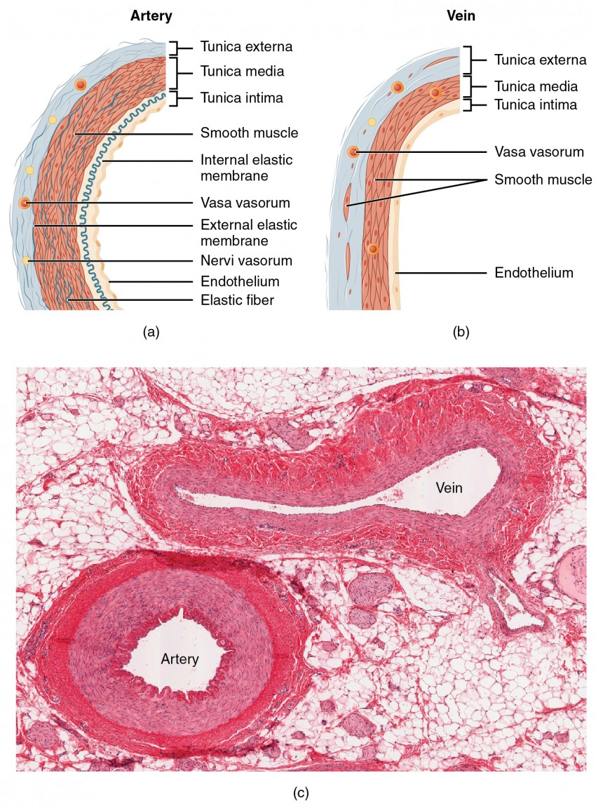
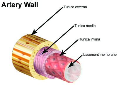
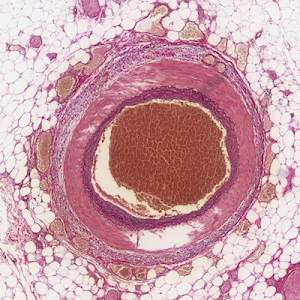
Tissue Of Arteries And Veins Under The Microscopic Physiology Of Histology Of Blood Vessels Artery And Vein Prepared Microscope Slide Mammal Artery Vein C S 7 M H E Microscope Slide Microscope Under The Microscope Artery And Vein Cs Travis Hale Mammal Artery And Vein Transverse Section 64x MammalsArtery under the Microscope Arteries are blood vessels that deliver oxygenrich blood from the heart to the tissues of the body Each artery is a muscular tube lined with smooth tissue that has three layers The intima is the inner layer lined by a smooth tissue called endothelium The media is a layer of muscle that lets arteries handle the high pressures from the heartUsually, arteries and veins are connected through a system of tiny blood vessels called capillaries As blood flows from arteries into capillaries, the blood pressure is lessened While in capillaries, the blood gives up oxygen and picks up waste products Then the blood flows from the capillaries into veins Abnormal Blood Flow in an AVM or DAVF


Histology Vascular


Histopathology Of Hypertensive Renal Disease Light Micrograph Stock Photo Image Of Chronic High
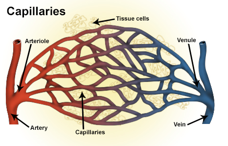
Photo under microscope, hypertony, hypertonic, vessel, artery, vein, capillary, high, blood Arteriovenous Graft An arteriovenous (AV) fistula is an abnormal connection between an artery and a vein Normally, blood flows from your arteries to yourCapillaries, the smallest and most numerous of the blood vessels, form the connection between the vessels that carry blood away from the heart (arteries) and the vessels that return blood to the heart (veins) The primary function of capillaries is the exchange of materials between the blood and tissue cells What is the structure of capillaryArtery Tissue Label Artery and Vein Slide Labeled Rabbit Capillaries 40X Magnification Arteries Veins Capillaries Comparison Plant Stem Cell Under Microscope Plant Cell Under Microscope
:background_color(FFFFFF):format(jpeg)/images/library/3610/5NHWYTWfF0zpaLrwavgwQ_Venule.png)


Blood Vessels Histology And Clinical Aspects Kenhub


Artery Vein And Capillary Cs Mammal Cross Sect Of 3 Vascular Tissues On One Slide H And E Histology Prepared Microscope Slides The Science Shop
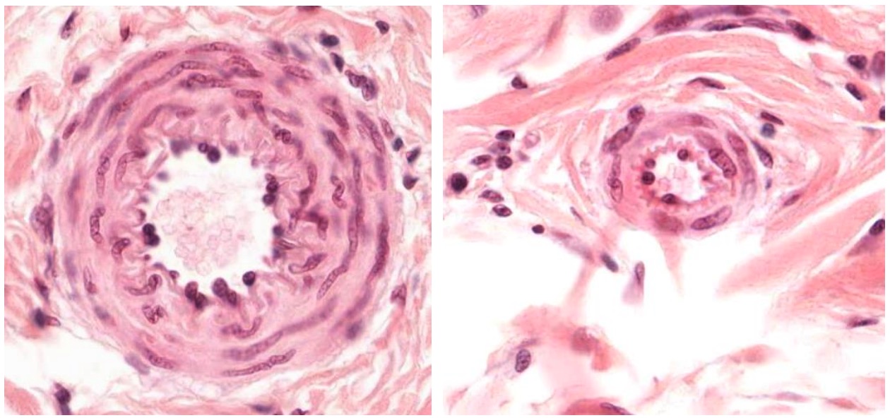
When we look at an artery and vein under a microscope we can see that there are differences between the On our video tutorial we will go through the histological features of arteries and veins This feature is not available right now The arteries smaller branches are referred to as arterioles and capillariesUnder the microscope, the lumen and the entire tunica intima of a vein will appear smooth, whereas those of an artery will normally appear wavy because of the partial constriction of the smooth muscle in the tunica media, the next layer of blood vessel wallsCapillary Microscopy Capillary microscopy is most useful as a prognostic factor in Raynaud's disease or mixed connective tissue disease, to differentiate 'undifferentiated' connective tissue disease to distinguish cutaneous lupus from dermatomyositis, and to confirm the diagnoses of systemic scleroderma, dermatomyositis, and systemic lupus erythematosus


27 Differences Between Arteries And Veins Arteries Vs Veins


Structure And Function Of Blood Vessels Anatomy And Physiology Ii
Arterioles carry blood and oxygen into the smallest blood vessels, the capillaries Capillaries are so small they can only be seen under a microscope The walls of the capillaries are permeable to oxygen and carbon dioxide Oxygen moves from the capillary toward theSection A 10% discount applies if you order more than 10 of this item and 15% discount applies if you order more than 25 of this item Triarch Incorporated offers superior prepared microscope slidesProcedure 1 Microscopy Examine prepared microscope slides of an artery, a capillary, and a vein The capillary may be on a separate slide Use colored pencils to draw what you see and label your diagrams with the listed terms


Exercise 21 Anatomy Of Blood Vessels Ppt Video Online Download
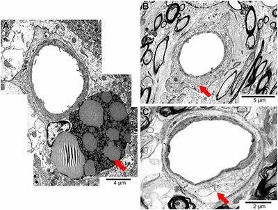


Frontiers Solving An Old Dogma Is It An Arteriole Or A Venule Frontiers In Aging Neuroscience
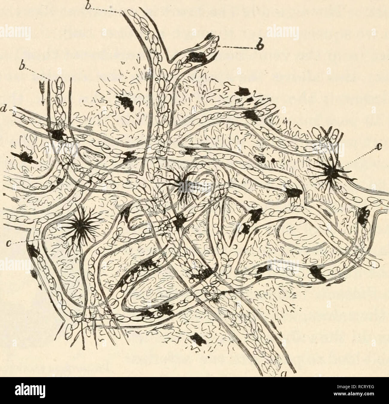
The average length of a capillary is 05 1 mm, average diameter, 8 µ or often less than a red cell, average velocity of flow, 05 1 mm per second Capillaries are lined by a layer of the flat endothelial cells bound together by an intracellular cement substance A basement membrane surrounds capillary endothelial cellsCapillaries are the thinnest blood vessels in the body These vessels are found throughout the body and bring oxygen and necessary nutrients to every cell in the body and remove cell waste Capillary walls are selectively permeable, allowing some, but not all substances throughDescription This slide shows artery, vein and capillaries in a transverse (cross) section, stained with H&E (haematoxylin and eosin) to show nuclei in blue and the extracellular matrix and cytoplasm in


Cardiac Muscle Ppt Video Online Download
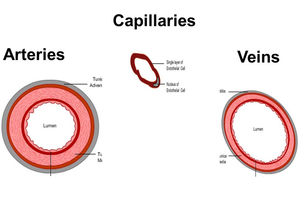


Topic Circulatory System Ppt Video Online Download
Microcirculation Microscope $ USD The Nailfold Capillaroscopy Microcirculation Microscope is an advance medical photoelectric apparatus,equipped with built in special LED light source,used mainly in observation on human nail fold capillary microcirculation or term as video Nailfold capillaroscopy, DescriptionArteries and veins consist of three layers an outer tunica externa, a middle tunica media, and an inner tunica intima Capillaries consist of a single layer of epithelial cells, the tunica intima (credit modification of work by NCI, NIH) This article has been modified from " Mammalian Heart and Blood Vessels ," by OpenStax, Biology, CC BY 40Endothelium Smooth muscle Connective tissue What does a small muscular artery and a vein look like under a microscope?


Cardiovascular


Biol 2402 Lab Ppt The Cardiovascular System Blood
Lungs The pulmonary arteries from right ventricle;Under the microscope, the lumen and the entire tunica intima of a vein will appear smooth, whereas those of an artery will normally appear wavy because of the partial constriction of the smooth muscle in the tunica media, the next layer of blood vessel walls Normally travels in arteries Vein ↑ A blood vessel which carries blood from the body to the heart Deoxygenated Blood ↑ Blood that contains low levels of oxygen This normally travels in veins Capillary ↑ A small blood vessel just 5–10 μm in


Blood Vessels Use A Microscope Slide Showing The Three Types Of Blood Vessels And Blood Vessel Models To Identify Each Of The Following Flashcards Quizlet


Bl V
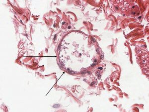
Under the microscope, capillaries are hard to find as they have only one layer of cells, endothelium, and are usually collapsed in histological sections Capillaries are exchange vessels, as the interchange of nutrients/waste between blood and organs takes place across their walls Veins A vein is a blood vessel that conducts blood toward the heart Veins have thinwalls, and compared to arteries,It's very simple Arteries are the vessels that carry your blood from the heart to the extremities of your body As they reach your extremities, such as your fingers, toes, earlobes, eyelids and of course all the organs within you body, includingOn the right is a fenestrated capillary


Circulatory System The Histology Guide


Structure And Function Of Blood Vessels Anatomy And Physiology Ii


Vessel Comparison Bioninja


Veins And Arteries Yr 9


Cross Section Of Artery Vein For Fl Youtube


Seer Training Classification Structure Of Blood Vessels


Vessel Comparison Bioninja


Capillary


Histopathology Of Hypertensive Renal Disease Light Micrograph Stock Photo Image Of Artery Vein


Circulatory System The Histology Guide


Histology Laboratory Manual


Blood Vessels Lab
:max_bytes(150000):strip_icc()/GettyImages-464612781-edc0c4a8ab20491580bc3dd7acd4f4d2.jpg)


Capillary Structure And Function In The Body


Cardiovascular


Histology Of Blood Vessels
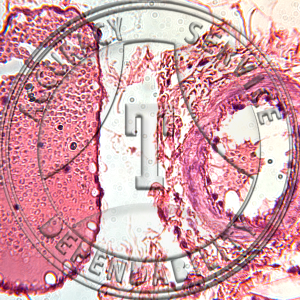


Artery Vein Capillary Prepared Microscope Slide


Blood Vessels Lab


Human Structure Virtual Microscopy


Artery Vein Nerve Elastic Tissue Stain Prepared Microscope Slide


Cardiovascular System Histology


1


Blood Vessels A Level Biology Student


Histology Of Blood Vessels


Seer Training Classification Structure Of Blood Vessels


Unit J Notes 1 Blood Vessels Handout Mr Lesiuk


Cardiovascular


Arteries


Human Structure Virtual Microscopy


Blood Vessels Under The Microscope Frontiers For Young Minds
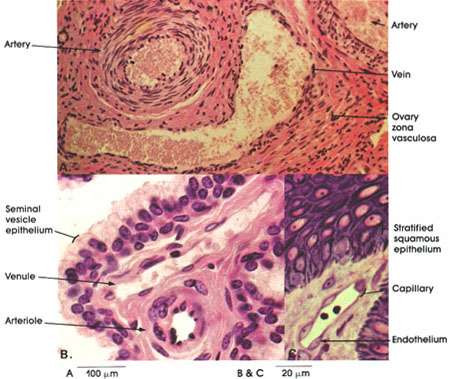


Anatomy Atlases Atlas Of Microscopic Anatomy Section 1 Cells
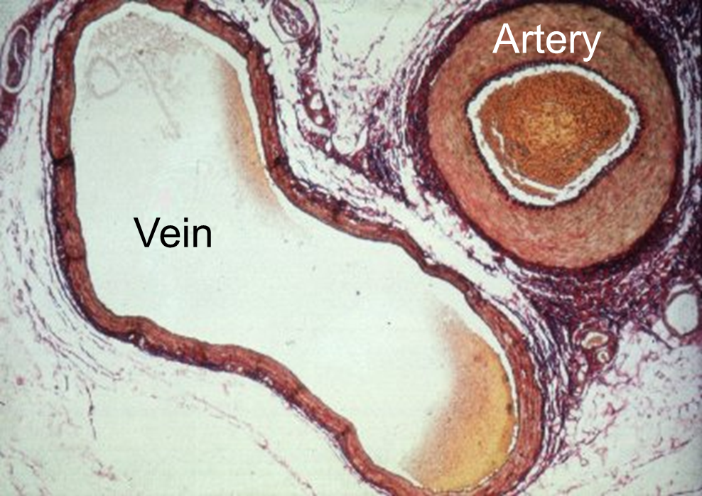


Cardiovascular System


A P Ii Exercise 19 Cardiovascular System Circulatory Pathways Lymphatic System Flashcards Quizlet


Histopathology Of Hypertensive Renal Disease Light Micrograph Stock Photo Image Of Capillary Blood


Figure 19 1a Generalized Structure Of Arteries Veins And Capillaries Artery A Ppt Video Online Download


Comparative Anatomy Of The Vasculature Of The Dog Canis Familiaris And Domestic Cat Felis Catus Paw Pad


Heart Veins Arteries Black And White Stock Photos Images Alamy


Chapter 10 Page 1 Histologyolm 4 0
:background_color(FFFFFF):format(jpeg)/images/library/3603/Tpmr9patPzlfw4MJozUjQ_Elastic_artery_-_Aorta__1_.png)


Blood Vessels Histology And Clinical Aspects Kenhub


Histopathology Of Hypertensive Renal Disease Light Micrograph Stock Image Image Of Micro Tissue


Untitled Document
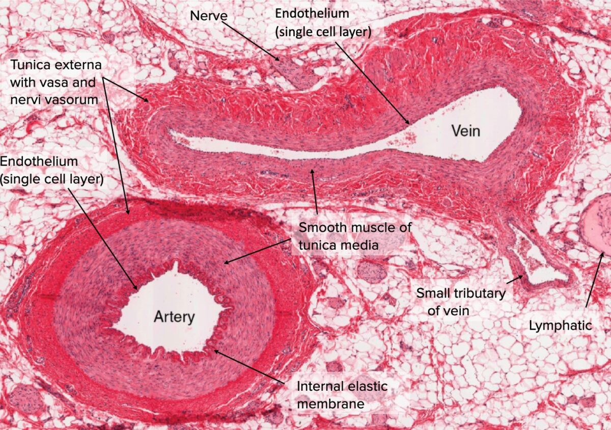


Arteries Concise Medical Knowledge


Cardiovascular System Histology


44 The Circulatory System Blood Vessels


Blood Vessels Arteries Capillaries More Video Lesson Transcript Study Com


Histopathology Of Hypertensive Renal Disease Light Micrograph Stock Image Image Of Disease Vessel
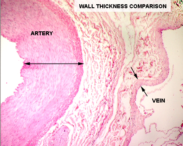


Exercise 12b


Activity 1 Examning The Microscopic Structure Of Arteries And Veins Flashcards Easy Notecards


Blood Vessels Lab


Histology Of Arteries Veins And Capillaries Preview Microscopic Anatomy Kenhub Youtube


Copyright C The Mcgraw Hill Companies Chapter 11 The Circulatory System The Circulatory System Introduction The Circulatory System Includes Both The Blood And Lymphatic Vascular Systems The Blood Vascular System Figure 11 1 Is Composed Of The


Embryology And Histology Of Bloodvessels


Blood Vessels Lab


Exercise 17 4 Histology Of The Blood Vessel Wall Chegg Com


Veins Arteries Microscope High Resolution Stock Photography And Images Alamy


Microscope World Blog Artery Under The Microscope


What Are The Main Characteristics Of The Arteries Veins And Capillaries Quora


Circulation Of Blood In The Human Body Through Arteries Veins And Capillaries Britannica


Veins Arteries Microscope High Resolution Stock Photography And Images Alamy


Topic 6 2 The Blood System Amazing World Of Science With Mr Green


Histolab3a Htm


Artery And Vein C S


Arteries Concise Medical Knowledge


Cardiovascular


Histology Of Blood Vessels


Cardiovascular


Ultrastructure Of Blood Vessels Arteries Veins Teachmeanatomy


1


Artery Vein Capillary Elastic Tissue Stain Prepared Microscope Slide


Cardiovascular System Histology


Elements Of Biology A Practical Text Book Correlating Botany Zoology And Human Physiology Biology 354 Human Physiology Trace The Artery Until It Breaks Out Into Very Tiny Tubes The Capillaries Notice That


Circulatory System The Histology Guide


Histopathology Of Hypertensive Renal Disease Light Micrograph Stock Photo Image Of Kidney Histology


Blood Vessels Lab


Blood Vessels Lab


Artery And Vein


Arteries


Vintage Microscope Slide Wards Artery Vein Capillaries C S 93 W 4043 007 Ebay


Blood Vessels Knowledge Amboss


No comments:
Post a Comment